You are here
Scientists 3D-print a ‘brain’ to learn the secret behind its folds
By Los Angeles Times (TNS) - Feb 09,2016 - Last updated at Feb 09,2016

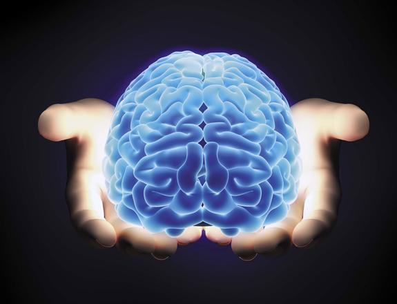
Photo courtesy of galleryhip.com
By 3D-printing a fake gel brain and watching it “grow”, scientists at Harvard University have discovered how the human cortex develops its creepy, classic folds.
The discovery, published in the journal Nature Physics, may solve a long-standing mystery about the structure of our grey matter and could even help shed light on certain disorders that may be linked to underfolding or overfolding of the brain.
The researchers “have provided the first experimental evidence of the theory of differential growth and demonstrated that physical forces — not just biochemical processes alone — play a critical role in neurodevelopment”, Ellen Kuhl of Stanford University, who was not involved in the study, wrote in a commentary. “Their findings could have far-reaching clinical consequences for diagnosing, treating and preventing a wide variety of neurological disorders.”
Think of the brain, and you might conjure up a pink, wrinkly object roughly the shape of a partly deflated basketball. But not every species’ brain has these telltale wrinkles — smaller animals such as rats have smooth, pink thinkers. Human foetuses don’t start developing these folds until about 23 weeks of gestation and don’t put the final touches on the branch-like network of creases until after they’re born.
Scientists have long known that the brain’s folded structure actually has some major benefits: It allows for far more connectivity across the cortex (the surface layer of our brain that consists of “grey matter”) than a smooth surface would.
“Each cortical neuron is connected to 7,000 other neurons, resulting in 0.15 quadrillion connections and more than 150,000km of nerve fibres,” Kuhl wrote.
But what causes these folds to develop has had scientists a little stumped. Many researchers have tried to identify the cellular or biochemical processes at work, but Lakshminarayanan Mahadevan, a physicist and applied mathematician at Harvard University, decided to study the physics of the structure itself, and develop a mathematical model of its behaviour.
“I have a long-standing interest in trying to understand how the body or bodies of animals organise themselves,” Mahadevan said. “I approach these problems from a mathematical perspective.”
Researchers have tried to get at this question for decades, Kuhl wrote. Roughly 40 years ago, another group of Harvard researchers suggested a physical model where the differences in growth within the brain’s tissues could explain fold formation.
“The model challenged the conventional wisdom that surface morphogenesis, pattern selection and evolution of shape are purely biological phenomena,” Kuhl wrote. “To no surprise, this rather hypothetical approach was perceived as highly controversial.”
Part of the problem was that there seemed to be no good way to answer this question, she added. Experiments with human brains can be “ethically questionable”, and experiments with rats or other small animals wouldn’t work because their brains are smooth. And usually, an experiment in non-living material wouldn’t show you how the brain develops these folds because non-living tissue doesn’t grow.
But for this paper, the scientists built a physical model that solved that last problem with some clever use of materials. They used magnetic resonance imagery from a smooth foetal brain at 22 weeks’ gestation and 3D-printed a cast to make a fake brain out of gel. This was the “white matter” of the brain, which they covered with a thin coat of rubbery gel to mimic the layer of “grey matter”, or cortical tissue.
The researchers then submerged the brain in a liquid solvent that caused that stretchy cortex-like layer to start growing. Sure enough, unnervingly brain-like folds began to emerge in the once-smooth surface.
Here’s what seems to be happening: The cortical tissue wants to keep growing but it’s anchored to the limited real estate of the white matter below it. As the cortex expands, that strain eventually causes the tissue to collapse, leading to the gyri (round features) and sulci (deep grooves) that cover the surface.
The next step, Mahadevan says, is to link these large-scale structural predictions to the process that may be contributing on a molecular level.
“In the end, all of them are related,” he said. “If I think about the shape of the folds in a foetal brain then yes, there are molecular processes: There are biochemical processes which cause cells to move, cause cells to divide, cause cells to change shape and cause cells to change in number.”
Ultimately, the research could help researchers better understand a variety of different neurological disorders, scientists said.
“Making these connections can help us identify topological markers for the early diagnosis of autism, schizophrenia or Alzheimer’s disease, and, ultimately, design more effective treatment strategies,” Kuhl said.
Related Articles
Damage to the brain’s outer layer caused by smoking may be reversible after quitting, but it could take years, a study said Tuesday.
PARIS — Personality traits such as moodiness or open-mindedness are linked to the shape of one’s brain, a study said on Wednesday.Researcher
British scientists have developed a new use for 3D printing, putting it to work to create personalised replica models of cancerous parts of the body to allow doctors to target tumours more precisely.


